Expression of the Fructose-1,6-Bisphosphatase 1 (FBP1) Gene in the Embryonic Bursa of Fabricius
Abstract
Background and Objective: Previous transcriptome analysis of isolated bursal B-cells at Embryonic Day (ED) 16 and ED19 revealed expression of the fructose-1,6-bisphosphatase 1 (FBP1) gene, which encodes a rate-limiting enzyme in the gluconeogenesis (GNG) pathway. To validate this finding, the goal of this project was to confirm FBP1 gene expression before and after the major B-cell differentiation event in the embryonic bursa.
Materials and Methods: Total RNA and frozen sections prepared from pooled bursas of ED15 and ED19 embryos were evaluated using quantitative polymerase chain reaction (qPCR) analysis and in situ hybridization (ISH).
Results: The qPCR results suggest that FBP1 gene expression decreases in the bursa during embryonic development. The ISH methodology localized FBP1 expression to the bursal follicles.
Conclusion: These results confirm the expression of the FBP1 gene during bursal B-cell development. We hypothesize that the expression of a rate-limiting enzyme in the GNG pathway confers metabolic flexibility on bursal B-cells.
Keywords:
Bursa of Fabricius, chicken embryo, fructose-1,6-bisphosphatase 1Article Metrics
License
Copyright (c) 2026 Gregory T. Pharr, Hui Wang, Nikhil Nuthalapati

This work is licensed under a Creative Commons Attribution 4.0 International License.
This is an open access article distributed under the terms of the Creative Commons Attribution License, which permits unrestricted use, distribution and reproduction in any medium, provided the original author and source are credited.
Article Citation Metrics
How to Cite




INTRODUCTION
Chicken B-cell development is guided by the primary lymphoid tissue microenvironment of the bursa of Fabricius1. The development of B-cells in the chicken initiates during the embryonic period, when the bursal mesenchyme is colonized by prebursal stem cells between embryonic days of development (ED) 9 and ED122. The stem cells are then recruited into the nascent bursal follicles and undergo proliferative expansion2. Within the developmental window of ED15 to ED17, a major B-cell differentiation event occurs that initiates the development of the pre-immune repertoire through immunoglobulin gene conversion3. The export of naïve B-cells commences by hatching and continues until bursal involution at sexual maturity4.
The long-term goal of our lab is to understand the microenvironmental factors responsible for the major B-cell differentiation event in the embryonic bursa. In a previous transcriptomics study of bursal B-cells isolated at ED16 and ED19, we identified the unexpected expression of a rate-limiting gluconeogenesis (GNG) enzyme, fructose-1,6-bisphosphatase 1 (FBP1)5. Therefore, the goal of this project was to validate the expression of FBP1 in the embryonic bursa as B-cells differentiate toward repertoire development.
MATERIALS AND METHODS
Experimental animals: Fertile eggs from the Ross 708 broiler parent stock were used in this project. Embryonated eggs were incubated at 99.5̊F and 60% relative humidity. This project was reviewed by the Mississippi State University Animal Care and Use Committee.
Bursa total RNA: Chicken embryos at ED15 and ED19 were euthanized by decapitation and total RNA was prepared from whole bursal tissue as described previously5. Two bursas were pooled for RNA preparation and this was defined as an experimental unit. Three experimental units from ED15 and three from ED19 were prepared and stored at -80̊C until analysis by qPCR.
Quantitative real-time PCR: Our qPCR procedure has been described previously6. Briefly, FBP1 gene expression levels were determined using the forward primer 5’-TTTTATGCT TGATCCGGCAATC-3’ and reverse primer 5’-ACCGCAGCATCA AAGTATTTAG-3’. For normalization to a housekeeping gene, the GAPDH forward primer 5’-TGCTGCCC AGAACATCATCC-3’ and reverse primer 5’-ACGGCAGGTCAGGTCAACAA-3’ were used. The reactions were performed in TaqPath qPCR Master Mix (No. A28525, Thermo Fisher, USA). The qPCR reactions were amplified using an AriaMX QPCR Systems thermocycler (Agilent Technologies, USA).
In situ hybridization: Chicken embryos at ED19 were euthanized by decapitation and whole bursas were embedded in optimum cutting temperature medium for the preparation of frozen sections. Based on tissue size, the cryomolds contained three bursas each. For ISH analysis, 10-µm-thick frozen sections were processed according to the manufacturer’s protocol using the RNAscope 2.5 HD Reagent Kit (No. 322371, Advanced Cell Diagnostics, USA). The negative control probe DapB (No. 310043) and positive control probe Gg-PPIB (No. 453371) were obtained from Advanced Cell Diagnostics, USA. The FBP1 gene probe covered nucleotide positions 24-1043 of the FBP1 gene coding sequence from accession number NM_001278048.2 in the National Center for Biotechnology Information.
RESULTS AND DISCUSSION
In a previous study identifying the genes expressed during embryonic bursal B-cell differentiation, transcripts from the FBP1 gene were detected and the read counts were two-fold higher in bursal B-cells before the major B-cell differentiation event5. The qPCR analysis in the present study also showed higher FBP1 gene transcript levels prior to bursal B-cell differentiation at ED15 in whole-tissue RNA (Fig. 1), confirming the previous gene expression analysis performed using gradient-isolated bursal B-cells from Hy-Line W-36 layer embryos.
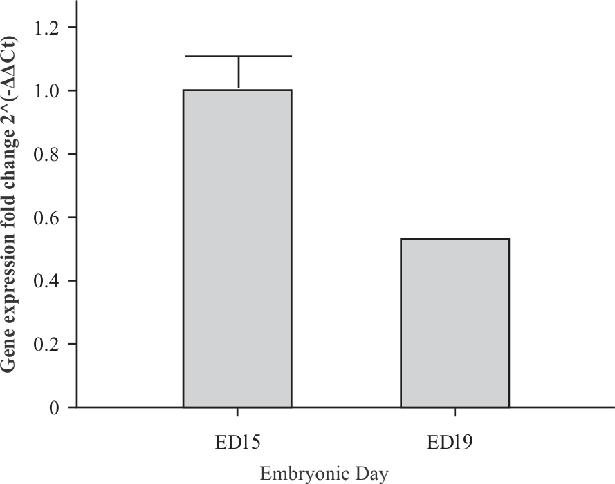
The bars stand for standard deviations. Three experimental units of ED15 and from ED19 were used in the analysis
To localize FBP1 expression in bursal tissue, ISH was conducted on tissue sections from the ED19 bursa because the follicles are more pronounced at this later stage of embryogenesis. Figure 2A shows an ED19 bursal frozen section hybridized with the control probe DapB, which represents the bacterial methylglyoxal synthase gene and, therefore, did not result in a signal in chicken tissue. The positive control probe Gg-PPIB, representing the chicken cyclophilin B gene, showed reactivity in the nascent follicles and epithelial lining of the bursal section, indicating that the tissue mRNA was intact (Fig. 2b). Reactivity with the FBP1 gene probe was confined to the nascent follicles (Fig. 2c). In conclusion, the ISH methodology, although not quantitative, showed that FBP1 gene expression was confined to the bursal follicles, supporting previous transcriptomic studies showing FBP1 gene transcripts in isolated embryonic bursal B-cells5.
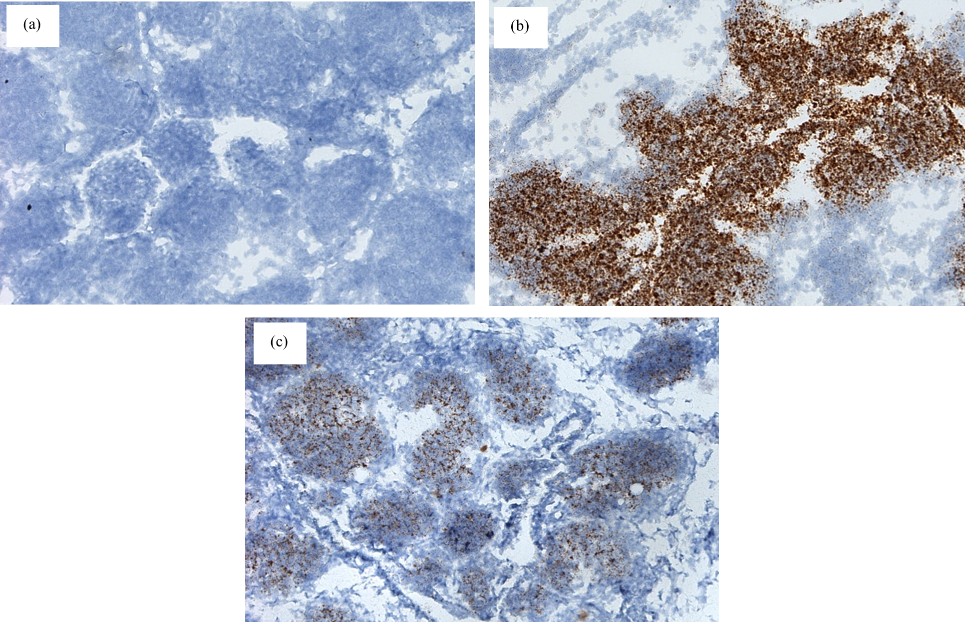
(a) Bursal tissue sections hybridized with the negative control probe DapB (10x magnification), (b) Bursal tissue sections hybridized with the positive control probe Gg-PPIB (10x magnification) and (c) Bursal tissue sections hybridized with the FBP1 gene probe (10x magnification)
The goal of this study was to validate the expression of the FBP1 gene in the embryonic bursa, as suggested by transcriptomic analysis. In liver tissues, FBP1 functions as a rate-limiting enzyme in GNG and catalyzes the conversion of fructose-1,6-bisphosphate into fructose-6-phosphate and inorganic phosphate7. In the chicken embryo, GNG primarily occurs in the liver and yolk sac membrane8. The expression of the FBP1 gene in a non-gluconeogenic tissue is difficult to explain at present but may reflect metabolic changes occurring during the last phase of embryonic development (ED15 to ED21)9. During this developmental phase, metabolic changes in the bursa also reflect the metabolism of the whole embryo10. Indeed, the seminal study by Rudrappa and Humphrey10 showed that, during the late embryonic period, bursal B-cells maintain energy production through β-oxidation and subsequently increase glycolysis and glutaminolysis toward hatching at ED21.
Taken together, the gene expression studies reported here and those of others10 suggest that bursal B-cells express genes encoding enzymes involved in both glycolysis and GNG. One possibility to explain this finding is that the expression of GNG enzymes could confer metabolic flexibility on bursal B-cells. Although, less well understood, it is plausible that differences in oxygen tension and nutrient availability may occur within the bursal follicles during the metabolic changes associated with the last phase of chick embryo development. Although the simultaneous occurrence of glycolysis and GNG, referred to as a futile cycle, has traditionally been considered to result in a loss of energy1, more recent studies suggest that metabolic futile cycles involving glycolysis and GNG could be active in different subcellular compartments within a cell11,12. Therefore, a futile cycle involving glycolysis and GNG could function to maintain an adequate supply of cellular substrates, which would be important in microenvironments with reduced nutrient availability12.
A second possibility is that the metabolic flexibility of bursal B-cells could be regulated by abbreviated GNG. In abbreviated GNG, only one of the three rate-limiting enzymes is expressed to provide substrates for the synthesis of macromolecules in rapidly proliferating cells in environments with reduced glucose availability13. A classic example of cellular utilization of abbreviated GNG is observed in cancer cells. Solid lung tumors show heterogeneity in oxygen and glucose concentrations throughout the tumor mass14. Protein expression studies revealed abbreviated GNG in cells mainly at the tumor margin, whereas cells in the hypoxic tumor center showed a predominantly glycolytic phenotype14. Importantly, cells in metastases from the primary tumor exhibited either an abbreviated GNG phenotype or a mixed glycolytic/abbreviated GNG phenotype, leading to the conclusion that the metabolic flexibility of cells with the mixed phenotype could facilitate adaptation to different microenvironments during metastasis to distant tissues14.
In summary, this study validates our previous transcriptomic work, which identified transcripts from the FBP1 gene in embryonic bursal B-cells5. Additional research will be required to determine the role of the FBP1 enzyme in bursal B-cell metabolism during embryonic B-cell development. Immediate future studies will be designed to assess the expression of the other two rate-limiting enzymes of GNG and the rate-limiting enzymes of glycolysis in bursal tissue sections and at the single-cell level, followed by metabolomic evaluation of isolated bursal B-cells.
CONCLUSION
The results of this project confirm the expression of FBP1 during the last phase of chick embryo development, which, to our knowledge, represents the first demonstration of the expression of a gene encoding a GNG enzyme in bursal B-cells.
ACKNOWLEDGMENT
This project was supported in part by an Agriculture and Food Research Initiative Program competitive grant (No. 2017-67016-26799) from the U.S. Department of Agriculture.